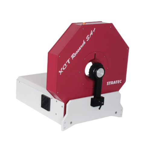
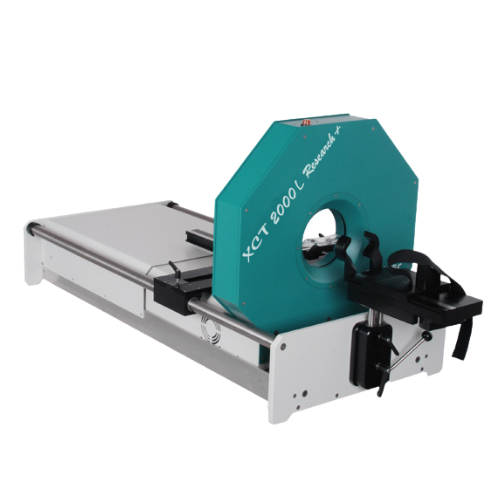
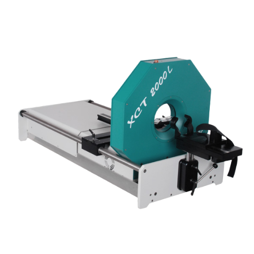
pQCT / xCT technology
Peripheral Quantitative Computed Tomography – pQCT
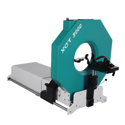
XCT 3000
pQCT – the third dimension in bone densitometry
With conventional area-projected bone densitometry techniques (DXA) bone strength cannot be determined adequately because they are lacking information about bone geometry.

With the pQCT technology bone strength with respect to bending, torsion, and compression can be calculated from bone’s cross sectional geometry. In addition the results are given as physically correct density units in g/cm³, whereas area projected techniques give only g/cm². Therefore the density values from the pQCT are independent from bone size. This is especially important for measurements on children. With the pQCT technology cortical and trabecular bone can be analysed separately and bone changes can be diagnosed reliably.
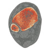
Additional morphometric parameter like endosteal and periosteal perimeter and bone cross sectional area are accessible in vivo. pQCT measurements at tibia or radius are performed in a wide range in clinical routine. In paediatrics the advantages of pQCT are especially distinct. It is the only method that allows to differentiate bone growth from other processes. In preclinical research specialised ultra high resolution scanner rare used in many pharmacological studies.
Relation between muscular demands and the resulting bone
But pQCT is even one step further than traditional methods: In addition to bone parameters it also quantifies muscle properties. This allows to quantify the relation between muscle and bone which is essential for the step from Osteoporosis diagnostics to diagnostics of Sarcopenia, Dynapenia and Frailty.
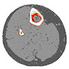
Essential for bone geometry and bone mass is the muscular demand on the bone. Julius Wolf postulated “bone geometry follows function” and Harold Frost specified the more detailed “Mechanostat” theorem. Therefore bone diagnostics must always be accompanied by a sufficient analysis of muscular properties and abilities. pQCT measurements (e.g. at 66% distal tibia) utilize this concept: it allows to quantify the relation between muscular demands and the resulting bone geometry and bone mass on the individual and site-specific level.

This combined analysis is essential to differentiate for example between a primary Osteoporosis and the effects of diuse or lack of mobility. According to Frosts Mechanostat both will result in loss of muscle mass. The difference can therefore not be found in the bone but in its combination with muscle properties and function. Therefore it is essential to quantify the site-specific relation between muscle and bone. In this context the cross-sectional area of the muscle can be used as a surrogate for peak muscular forces, one of the key osteo-anabol parameters. The examples on the right show on the top a healthy muscle-bone relation, in the middle a primary osteoporosis and on the bottom the long-term effects of a spinal cord injury (SCI).

In addition, newest research results show that especially for Sarcopenia, Dynapenia and Frailty the parameter of muscle density (specific property of muscle tissue) in addition to the muscle geometry is essential. The reason for this is currently considered to be mainly the inter- and intra-muscular fatty infiltration. pQCT allowing the combination of geometry and density measurement at an extremely low cost of radiation is therefore the diagnostic method of choice. To add even more details the pQCT based quantification of muscle and bone can be extended by the quantification of muscle function and performance based on the Leonardo Mechanography systems – for example using the complete pQCT Bone & Muscle Bundle.


